Directory:LASIK Center
| LASIK Center | |
 | |
| Slogan | LASIK Resources for a Clearer Future |
|---|---|
| Type | [[Company_Type:=Private|Private]] |
| Founded | [[Year_Started:=2007|2007]] |
| Founder | Garrett Minks |
| Headquarters | Template:Country data US [[City:=Houston|Houston]], [[State_Name:=Texas|Texas]], [[Country_Name:=United States|USA]] |
| Key people | [[Key_Person1:=Garrett Minks|Garrett Minks]], CEO |
| Industry | LASIK Help |
| Revenue | |
| Operating income | |
| Net income | |
| Employees | 1 (2007) |
| Owner | Garrett Minks |
| Contact | LASIK Center 22415 Water Edge Lane Katy TX US 77494 832.643.8266 [mailto:garrettminks@gmail.com Email] |
| Reference | Year End: 12/31 NAICS:51611 Entity: [[Entity_Type:=Sole proprietor|Sole]] |
LASIK Center (short for Laser-Assisted in Situ Keratomileusis center) is the definitive listing on the internet for all of your LASIK eye surgery needs. If you would like to advertise with the LASIK Center and <adsense>
google_ad_client = "pub-3695520715701375";
google_ad_output = "textlink";
google_ad_format = "ref_text";
google_cpa_choice = "CAAQhOSQ_QEaCJ0mn-838cQbKKjntoQB";
google_ad_channel = "7158149505";
</adsense>
Resources
Google has systematically picked the best LASIK services on the web.
<adsense> google_ad_client = "pub-3695520715701375"; google_ad_width = 336; google_ad_height = 280; google_ad_format = "336x280_as"; google_ad_type = "text_image"; //2007-03-06: Centiare google_ad_channel = "0332613634+8099356366"; google_color_border = "336699"; google_color_bg = "FFFFFF"; google_color_link = "0000FF"; google_color_text = "000000"; google_color_url = "008000"; </adsense>
Procedure
Preoperative
Patients wearing soft contact lenses typically are instructed to stop wearing them approximately 7 to 10 days before surgery. One industry body recommends that patients wearing hard contact lenses should stop wearing them for a minimum of six weeks plus another six weeks for every three years the hard contacts had been worn. Before the surgery, the patient's corneas are examined with a pachymeter to determine their thickness, and with a topographer to measure their surface contour. Using low-power lasers, a topographer creates a topographic map of the cornea. This process also detects astigmatism and other irregularities in the shape of the cornea. Using this information, the surgeon calculates the amount and locations of corneal tissue to be removed during the operation. The patient typically is prescribed an antibiotic to start taking beforehand, to minimize the risk of infection after the procedure.
Operation
The operation is performed with the patient awake and mobile; however, the patient typically is given a mild sedative (such as Valium) and anesthetic eye drops.
LASIK is performed in two steps. The first step is to create a flap of corneal tissue. A corneal suction ring is applied to the eye, holding the eye in place. The step in the procedure can sometimes cause small blood vessels to burst, resulting in bleeding or subconjunctival hemorrhage into the white (sclera) of the eye, a harmless side effect that resolves within several weeks. Increased suction typically causes a transient dimming of vision in the treated eye. Once the eye is immobilized, the flap is created. This process is achieved with a mechanical microkeratome using a metal blade, or a femtosecond laser microkeratome (procedure known as IntraLASIK) that creates a series of tiny closely arranged bubbles within the cornea.[3] A hinge is left at one end of this flap. The flap is folded back, revealing the stroma, the middle section of the cornea. The process of lifting and folding back the flap can be uncomfortable.
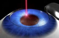
The second step of the procedure is to use an excimer laser (193 nm) to remodel the corneal stroma. The laser vaporizes tissue in a finely controlled manner without damaging adjacent stroma by releasing the molecular bonds that hold the cells together. No burning with heat or actual cutting is required to ablate the tissue. The layers of tissue removed are tens of micrometers thick. Performing the laser ablation in the deeper corneal stroma typically provides for more rapid visual recovery and less pain.
During the second step, the patient's vision will become very blurry once the flap is lifted. He/she will be able to see only white light surrounding the orange light of the laser. This can be disorienting.
Currently manufactured excimer lasers use an eye tracking system that follows the patient's eye position up to 4,000 times per second, redirecting laser pulses for precise placement within the treatment zone. The energy of each pulse is usually in the milliwatt range Typically, each pulse is on the order of 10–20 nanoseconds. After the laser has reshaped the stromal layer, the LASIK flap is carefully repositioned over the treatment area by the surgeon, and checked for the presence of air bubbles, debris, and proper fit on the eye. The flap remains in position by natural adhesion until healing is completed.
Postoperative
Patients are usually given a course of antibiotic and anti-inflammatory eye drops. These are discontinued in the weeks following surgery. Patients are also given a darkened pair of goggles to protect their eyes from bright lights and protective shields to prevent rubbing of the eyes when asleep.
Patient satisfaction
Various surveys have been performed to determine patient satisfaction with LASIK. These surveys have found most patients to be satisfied, with anywhere from 92–98% of respondents describing themselves as satisfied.[1][2][3]
Should I be Worried about Complications?
In 2003, the Medical Defence Union (MDU), the largest insurer for doctors in the United Kingdom, reported a 166% increase in claims involving laser eye surgery; however, the MDU averred that these claims resulted primarily from patients' “unrealistic expectations” of LASIK rather than “faulty surgery”.[4] A 2003 study reported in the medical journal Ophthalmology found that nearly 18% of treated patients and 12% of treated eyes needed retreatment.[5] The authors concluded that “higher initial corrections, astigmatism, and older age are risk factors for LASIK retreatment.”
On October 10, 2006, WebMD reported that a statistical analysis revealed the risks of infection due to contact lens wear is greater than the risk of infection from LASIK.[6] Daily contact lens wearers have about a one in 100 chance of developing a serious lens-related eye infection over 30 years of use, and a one in 2,000 chance of suffering significant vision loss as a result. The researchers calculated the risk of significant vision loss due to LASIK surgery to be closer to one in 10,000 cases.
The incidence of refractive surgery patients having unresolved complications six months after surgery has been estimated from 3%[7] to 6%.[8] The following are some of the more frequently reported complications of LASIK[7][9]
- Overcorrection or undercorrection
- Halos or starbursts around light sources at night
- Light sensitivity
- Ghosts or double vision
- Wrinkles in flap
- Debris or growth under flap
- Thin or buttonhole flap
- Induced astigmatism
- Posterior vitreous detachment[10]
- Macular hole[11]
Sources
- ^ Saragoussi D, Saragoussi JJ. "[Lasik, PRK and quality of vision: a study of prognostic factors and a satisfaction survey.]" J Fr Ophtalmol. 2004 Sep;27(7):755-64. PMID 15499272.
- ^ Bailey MD, Mitchell GL, Dhaliwal DK, Boxer Wachler BS, Zadnik K. "Patient satisfaction and visual symptoms after laser in situ keratomileusis." Ophthalmology. 2003 Jul;110(7):1371–8. PMID 12867394.
- ^ McGhee CN, Craig JP, Sachdev N, Weed KH, Brown AD. "Functional, psychological, and satisfaction outcomes of laser in situ keratomileusis for high myopia." J Cataract Refract Surg. 2000 Apr;26(4):497–509. PMID 10771222.
- ^ http://news.bbc.co.uk/2/hi/health/2937512.stm
- ^ http://www.ncbi.nlm.nih.gov/entrez/query.fcgi?cmd=Retrieve&db=pubmed&dopt=Abstract&list_uids=12689897&query_hl=5
- ^ http://www.webmd.com/content/article/128/117072.htm
- ^ a b Council for Refractive Surgery Quality Assurance. "The most common complications of refractive surgery.". ComplicatedEyes.org.
- ^ Albietz JM, Lenton LM, McLennan SG. "Dry eye after LASIK: comparison of outcomes for Asian and Caucasian eyes." Clin Exp Optom. 2005 Mar;88(2):89–96.
- ^ [1]:
- Dry eyes
- ^ Mirshahi A, Schopfer D, Gerhardt D, Terzi E, Kasper T, Kohnen T. "Incidence of posterior vitreous detachment after laser in situ keratomileusis." Graefes Arch Clin Exp Ophthalmol. 2006 Feb;244(2):149-53. Epub 2005 Jul 26. PMID 16044328.
- ^ Arevalo JF, Mendoza AJ, Velez-Vazquez W, Rodriguez FJ, Rodriguez A, Rosales-Meneses JL, Yepez JB, Ramirez E, Dessouki A, Chan CK, Mittra RA, Ramsay RC, Garcia RA, Ruiz-Moreno JM. "Full-thickness macular hole after LASIK for the correction of myopia." Ophthalmology. 2005 Jul;112(7):1207–12. PMID 15921746.